Moya-Sáez E, Menchón-Lara RM, Sánchez-González J,
et al. K-CC-MoCo: A Fast k-Space-Based Respiratory Motion Correction for Highly Accelerated First-Pass Perfusion Cardiovascular MR.
Magn Reson Med. 95(6):3536-3549 (2026).
https://doi.org/10.1002/mrm.70287
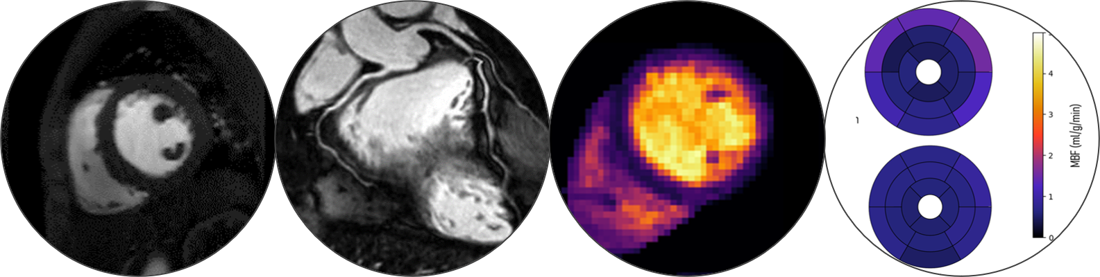
Ferrazzi G, Galan-arriola C, Velasco-Jimeno C
et al. Towards whole-heart quantitative myocardial perfusion using a dual-sequence framework with multiband acceleration (2025).
https://doi.org/10.1101/2025.08.20.666501
Olausson TE, Terpstra ML, Huttinga NRF,
et al. Free-running time-resolved first-pass myocardial perfusion using a multi-scale dynamics decomposition: CMR-MOTUS.
MAGMA (2025).
https://doi.org/10.1007/s10334-025-01291-x
Carvalho C, Gaspar A, Real C,
et al. Quantitative First-Pass Perfusion CMR: from technical principles to clinical practice (2025).
https://arxiv.org/abs/2502.13084
Silva SC, Correia TM, Manchado M, Power DM. Metamorphosis-associated immune system maturation in Senegalese sole.
Gen Comp Endocrinol, 369, 114755 (2025).
https://doi.org/10.1016/j.ygcen.2025.114755
Vitali M, Candeo A, Farina A,
et al. Accelerated dynamic lightsheet microscopy: unifying time-varying patterned illumination and low-rank and sparsity-constrained reconstruction.
Journal of Physics: Photonics, 7, 025005 (2025).
https://doi.org/10.1088/2515-7647/adad23
Obando M, Bassi A, Ducros N,
et al. Model-based deep learning framework for accelerated optical projection tomography.
Scientific Reports, 13, 21735 (2023).
https://doi.org/10.1038/s41598-023-47650-3
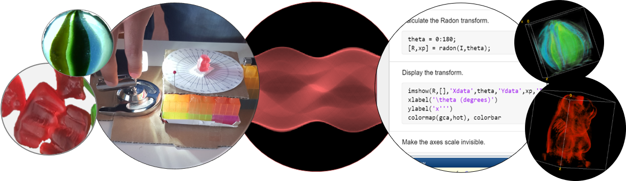
R Félix, D Palecek, T Correia. Colour science with lasers, gummy bears, and rainbows.
Science in School em> TEACH 66 (2024)
https://doi.org/10.5281/zenodo.10609154
Ismail TF, Strugnell W, Coletti C,
et al. Cardiac MR: From Theory to Practice
Front Cardiovasc Med 9, 826283 (2022).
https://doi.org/10.3389/fcvm.2022.826283
Tourais J, Scannell C, Schneider T
et al. High-Resolution Free-Breathing Quantitative First-Pass Perfusion Cardiac MR Using Dual-Echo Dixon With Spatio-Temporal Acceleration
Front Cardiovasc Med 9, 884221 (2022).
https://doi.org/10.3389/fcvm.2022.884221
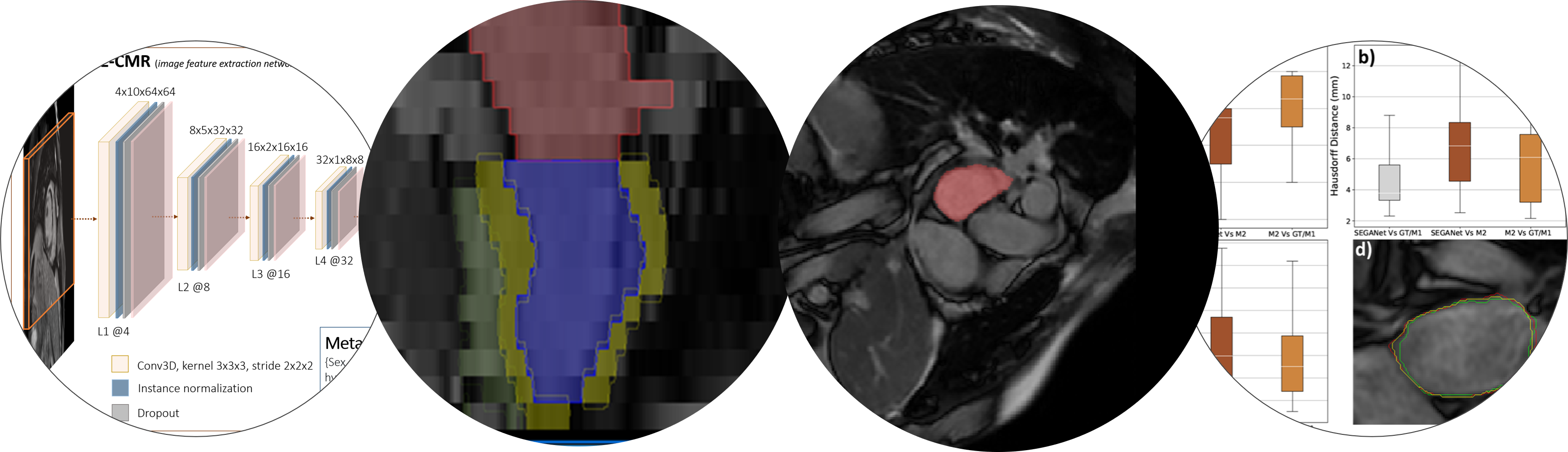
Lalande A, Chen Z, Pommier T
et al. Deep learning methods for automatic evaluation of delayed enhancement-MRI. The results of the EMIDEC challenge
Med Image Anal 79, 102428 (2022).
https://doi.org/10.1016/j.media.2022.102428
Martín-González E, Alskaf E, Chiribiri A
et al. Physics-Informed Self-supervised Deep Learning Reconstruction for Accelerated First-Pass Perfusion Cardiac MRI. In: Haq N., Johnson P., Maier A., Würfl T., Yoo J. (eds) Machine Learning for Medical Image Reconstruction. MLMIR 2021. Lecture Notes in Computer Science, vol 12964. Springer, Cham.
https://doi.org/10.1007/978-3-030-88552-6_9
Lourenço A, Kerfoot E, Grigorescu I
et al. Automatic Myocardial Disease Prediction from Delayed-Enhancement Cardiac MRI and Clinical Information. In: Puyol Anton E.
et al. (eds) Statistical Atlases and Computational Models of the Heart. M&Ms and EMIDEC Challenges. STACOM 2020.
Lecture Notes in Computer Science, vol 12592. Springer (2021).
https://doi.org/10.1007/978-3-030-68107-4_34
Lourenço A, Kerfood E, Dibblin C
et al. Left Atrial Ejection Fraction Estimation Using SEGANet for Fully Automated Segmentation of CINE MRI. In: Puyol Anton E.
et al. (eds) Statistical Atlases and Computational Models of the Heart. M&Ms and EMIDEC Challenges. STACOM 2020.
Lecture Notes in Computer Science, vol 12592. Springer (2021).
https://doi.org/10.1007/978-3-030-68107-4_14
Correia T, Ginami G, Rashid I
et al. Accelerated high-resolution free-breathing 3D whole-heart T2-prepared black-blood and bright-blood cardiovascular magnetic resonance.
J Cardiovasc Magn Reson 22, 88 (2020).
https://doi.org/10.1186/s12968-020-00691-3
Bustin A, Rashid I, Cruz G
et al. 3D whole-heart isotropic sub-millimeter resolution coronary magnetic resonance angiography with non-rigid motion-compensated PROST.
J Cardiovasc Magn Reson 22, 24 (2020).
https://doi.org/10.1186/s12968-020-00611-5
Varela M, Anjari M, Correia T
et al. High-resolution CINE MRI allows estimation of 3D regional atrial strains.
European Heart Journal, Volume 41, Issue Supplement_2, ehaa946.0244 (2020).
https://doi.org/10.1093/ehjci/ehaa946.0244
Lourenço A, E Kerfoot E, Dibblin C
et al. Automatic estimation of left atrial function from short-axis CINE-MRI using machine learning.
European Heart Journal, Volume 41, Issue Supplement_2, ehaa946.0229 (2020).
https://doi.org/10.1093/ehjci/ehaa946.0229
Scannell CM, Correia T, Villa ADM
et al. Feasibility of free-breathing quantitative myocardial perfusion using multi-echo Dixon magnetic resonance imaging.
Sci Rep 10, 12684 (2020).
https://doi.org/10.1038/s41598-020-69747-9
Varela M, Queiros S, Anjari M
et al. Strain maps of the left atrium imaged with a novel high-resolution CINE MRI protocol. Annual International Conference of the IEEE Engineering in Medicine and Biology Society.
IEEE Engineering in Medicine and Biology Society 2020 Jul;2020:1178-1181 (2020).
https://doi.org/10.1109/embc44109.2020.9175383
Nordio G, Schneider T, Cruz G,
et al. Whole‐heart T1 mapping using a 2D fat image navigator for respiratory motion compensation.
Magn Reson Med; 83: 178– 187 (2020).
https://doi.org/10.1002/mrm.27919
Velasco Forte MN, Valverde I, Prabhu N
et al. Visualization of coronary arteries in paediatric patients using whole-heart coronary magnetic resonance angiography: comparison of image-navigation and the standard approach for respiratory motion compensation.
J Cardiovasc Magn Reson 21, 13 (2019).
https://doi.org/10.1186/s12968-019-0525-8
Correia T, Schneider T, Chiribiri. A Model-Based Reconstruction for Highly Accelerated First-Pass Perfusion Cardiac MRI. In: Shen D.
et al. (eds) Medical Image Computing and Computer Assisted Intervention – MICCAI 2019. MICCAI 2019.
Lecture Notes in Computer Science, vol 11765. Springer (2019).
https://doi.org/10.1007/978-3-030-32245-8_57
Davis SP, Kumar S, Wisniewski L
et al. Exploiting patterned illumination and detection in optical projection tomography. Proc. SPIE BIOS 10883, Three-Dimensional and Multidimensional Microscopy: Image Acquisition and Processing XXVI, 108830M (2019).
https://doi.org/10.1117/12.2508653
Bustin A, Ginami G, Cruz G
et al. Five‐minute whole‐heart coronary MRA with sub‐millimeter isotropic resolution, 100% respiratory scan efficiency, and 3D‐PROST reconstruction.
Magn Reson Med 81: 102– 115 (2018).
https://doi.org/10.1002/mrm.27354
Correia T, Ginami G, Cruz G
et al. Optimized respiratory‐resolved motion‐compensated 3D Cartesian coronary MR angiography.
Magn Reson Med 80: 2618– 2629 (2018).
https://doi.org/10.1002/mrm.27208
Davis SP, Wisniewski L, Kumar S
et al. Slice-illuminated optical projection tomography.
Opt Lett 43, 5555-5558 (2018).
https://doi.org/10.1364/OL.43.005555
Correia T, Cruz G, Schneider T
et al. Accelerated nonrigid motion‐compensated isotropic 3D coronary MR angiography.
Med Phys 45: 214-222 (2018).
https://doi.org/10.1002/mp.12663
Ducros N, Correia T, Bassi A
et al. Reconstruction of an optical inhomogeneity map improves fluorescence diffuse optical tomography.
Biomedical Physics & Engineering Express 2(5): 055020 (2016).
https://doi.org/10.1088/2057-1976/2/5/055020
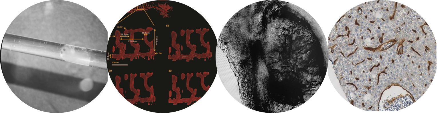
Kumar S, Lockwood N, Ramel M
et al. Quantitative in vivo optical tomography of cancer progression & vasculature development in adult zebrafish.
Oncotarget 7: 43939-43948 (2016).
https://doi.org/10.18632/oncotarget.9756
Kumar S, Lockwood N, Ramel M
et al. In vivo multiplexed OPT and FLIM OPT of an adult zebrafish cancer disease model. in
Biomedical Optics 2016, OSA Technical Digest (Optical Society of America), paper CTu4A.2 (2016).
https://doi.org/10.1364/CANCER.2016.CTu4A.2
Correia T, Koch M, Ale A
et al. Patch-based anisotropic diffusion scheme for fluorescence diffuse optical tomography—part 2: image reconstruction.
Physics in Medicine and Biology 61(4): 1439-1451 (2016).
https://doi.org/10.1088/0031-9155/61/4/1452
Correia T, Arridge S. Patch-based anisotropic diffusion scheme for fluorescence diffuse optical tomography—part 1: technical principles.
Physics in Medicine and Biology 61(4): 1452-1475 (2016).
https://doi.org/10.1088/0031-9155/61/4/1439
Correia T, Lockwood N, Kumar S
et al. Accelerated Optical Projection Tomography Applied to In Vivo Imaging of Zebrafish.
PLOS ONE 10(8): e0136213 (2015).
https://doi.org/10.1371/journal.pone.0136213
Kumar S, Lockwood N, Andrews N
et al. Rapid in-vivo Optical Projection Tomography of Larval and Adult Zebrafish Disease Models with Angular Multiplexing and FLIM-FRET. in
Optics in the Life Sciences, OSA Technical Digest (Optical Society of America), paper OT2D.2 (2015).
https://doi.org/10.1364/BODA.2015.OT2D.2
Dempsey L, Cooper R, Roque T
et al. Data-driven approach to optimum wavelength selection for diffuse optical imaging.
J Biomed Opt 20(1) 016003 (2015).
https://doi.org/10.1117/1.JBO.20.1.016003
V Soloviev, T Correia, S Arridge. Fluorescence lifetime measurements in turbid media. in Fluorescence Lifetime Spectroscopy and Imaging for tissue biomedical diagnostics, ed. by L Marcu, P French, D Elson, 283-322, CRC Press (2014).
https://doi.org/10.1201/b17018
Robertson JL, Ghosh A, Correia T
et al. Effect of Blood in the Cerebrospinal Fluid on the Accuracy of Cerebral Oxygenation Measured by Near Infrared Spectroscopy. In: Swartz H.M., Harrison D.K., Bruley D.F. (eds) Oxygen Transport to Tissue XXXVI.
Advances in Experimental Medicine and Biology, vol 812. Springer, New York, NY (2014).
https://doi.org/10.1007/978-1-4939-0620-8_31
Correia T, Rudge T, Koch M
et al. Wavelet-based data and solution compression for efficient image reconstruction in fluorescence diffuse optical tomography.
J Biomed Opt 18(8) 086008 (2013).
https://doi.org/10.1117/1.JBO.18.8.086008
Chamorro-Servent J, Abascal J, Aguirre J
et al. Use of Split Bregman denoising for iterative reconstruction in fluorescence diffuse optical tomography.
J Biomed Opt 18(7) 076016 (2013).
https://doi.org/10.1117/1.JBO.18.7.076016
Correia T, Ducros N, D’Andrea C
et al. Quantitative fluorescence diffuse optical tomography in the presence of heterogeneities.
Opt Lett 38, 1903-1905 (2013).
https://doi.org/10.1364/OL.38.001903
Correia T, Rudge T, Arridge S. Efficient image reconstruction in fluorescence diffuse optical tomography (fDOT) using data and solution compression. in Diffuse Optical Imaging IV, P. Taroni and H. Dehghani, eds., Vol. 8799 of SPIE Proceedings (Optical Society of America), paper 87990H (2013).
https://doi.org/10.1117/12.2032548
Papademetriou MD, Richards J, Correia T et al. Cortical Mapping of 3D Optical Topography in Infants. In: Van Huffel S., Naulaers G., Caicedo A., Bruley D.F., Harrison D.K. (eds) Oxygen Transport to Tissue XXXV. Advances in Experimental Medicine and Biology, vol 789. Springer, New York, NY (2013). https://doi.org/10.1007/978-1-4614-7411-1_61
Soloviev V, Zacharakis G, Spiliopoulos G et al. Tomographic imaging with polarized light. J. Opt. Soc. Am. A 29, 980-988 (2012). https://doi.org/10.1364/JOSAA.29.000980
Correia T, Lloyd-Fox S, Everdell N et al. Three-dimensional optical topography of brain activity in infants watching videos of human movement. Physics in Medicine and Biology 57(5): 1135-1146 (2012). https://doi.org/10.1088/0031-9155/57/5/1135
Abascal JFP‐J, Chamorro‐Servent J, Aguirre J et al. Fluorescence diffuse optical tomography using the split Bregman method. Med. Phys., 38: 6275-6284 (2011). https://doi.org/10.1118/1.3656063
Correia T, Aguirre J, Sisniega A et al. Split operator method for fluorescence diffuse optical tomography using anisotropic diffusion regularisation with prior anatomical information. Biomed. Opt. Express 2, 2632-2648 (2011). https://doi.org/10.1364/BOE.2.002632
Correia T, Gibson A, Hebden J. Identification of the optimal wavelengths for optical topography: a photon measurement density function analysis. J. Biomed. Opt. 15(5) 056002 (2010). https://doi.org/10.1117/1.3484747
Correia T, Banga A, Everdell N et al. A quantitative assessment of the depth sensitivity of an optical topography system using a solid dynamic tissue-phantom. Physics in Medicine and Biology 54(20): 6277-6286 (2009). https://doi.org/10.1088/0031-9155/54/20/016
Correia T, Gibson A, Schweiger M, Hebden J. Selection of regularization parameter for optical topography. J. Biomed. Opt. 14(3) 034044 (2009). https://doi.org/10.1117/1.3156839
Correia T, Gibson A, Hebden J. Identification of the optimal wavelengths in optical topography using photon density measurement functions. Proc. SPIE 7187, Biomedical Applications of Light Scattering III, 718718 (2009). https://doi.org/10.1117/12.809295
Hebden J, Correia T, Khakoo I et al. A dynamic optical imaging phantom based on an array of semiconductor diodes. Physics in Medicine and Biology 53(21): N407-N413 (2008). https://doi.org/10.1088/0031-9155/53/21/N01
Correia T, Gibson A, Hebden J. Optimal Selection of the Regularization Parameter for Optical Topography Image Reconstruction. in Biomedical Optics, OSA Technical Digest (CD) (Optical Society of America), paper BSuE49 (2008).https://doi.org/10.1364/BIOMED.2008.BSuE49
Hebden J, Bunker J, Correia T et al. An electrically-activated dynamic tissue-equivalent phantom for assessment of diffuse optical imaging systems. Physics in Medicine and Biology 53(2): 329-337 (2007). https://doi.org/10.1088/0031-9155/53/2/002